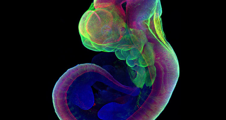
The new method developed at the Weizmann Institute allows biologists to learn better than ever how mammals develop in the early stages of gestation.
By Batya Jerenberg, World Israel News
Weizmann Institute of Science biologists have succeeded in growing mice outside of a uterus for longer than ever before, in a breakthrough that could help speed up research and cut the need for using live animals in lab experiments, as published Wednesday in the scientific journal Nature.
“It looks very spectacular,” Max Planck Institute for Molecular Genetics developmental biologist Alexander Meissner, who was not part of the team, told Science Magazine. “The fact that [the researchers] can culture these embryos and keep them alive for such a long time — it’s amazing.”
The record for keeping an embryo alive in a test tube was less than a week. Prof. Jacob Hanna of the Institute’s Molecular Genetics Department has been working for seven years with his group to extend that time. They have now developed a two-step process that enables them to grow mice for 11 days, over half-way through the 20-day gestation period.
That gave the scientists enough time to observe how the limbs are defined and organs formed.
“To us, that is the most mysterious and the most interesting part of embryonic development. Now, using this new method, we can look at these steps in detail and perform various experiments that weren’t possible before,” Hanna said in a statement released by the Institute.
The first step consisted of extracting embryos from pregnant mice before they implanted in the wall of the uterus, when they consisted only of spheres of 250 stem cells. They succeeded in attaching the microscopic balls instead to a special growth medium on a petri dish, where over the next two days they differentiated into three layers from which organs are formed.
Once they reached this stage, the embryos were put into test tubes filled with a nutrient solution. The beakers were placed on rollers that gently and continuously turned and moved them to keep the embryos bathed in the solution. The team also controlled the oxygen and carbon dioxide levels and the gas pressure inside the tubes to mimic the conditions inside a natural womb.
The scientists genetically tagged certain cells so they could follow organs’ development in fluorescent color. They even successfully incorporated human neural cells into the mice brains.
These procedures can help scientists of the future understand how normal – and abnormal – growth occurs in utero. The new method could be used to show the results of altering the conditions in the womb, or the effects of a virus infecting a fetus, Hanna said. The team hopes to also use their discovery to help doctors understand why so many pregnancies fail to implant and why developmental disorders occur, which could help the medical field enormously in the years to come.


